MGD-1k Dataset

Dataset overview

MGD-1k Dataset

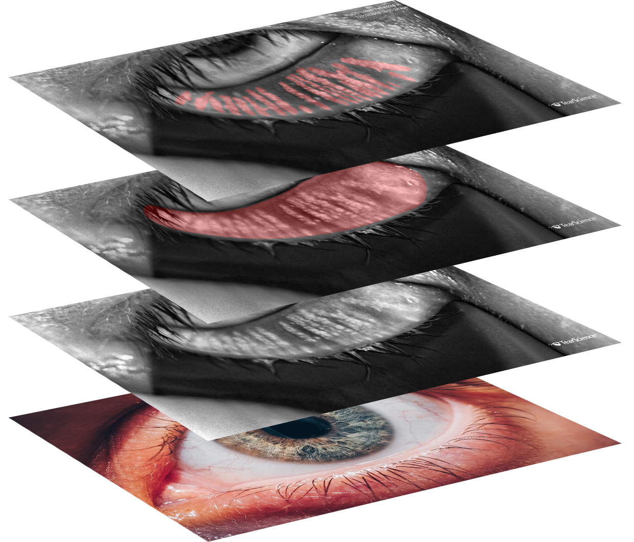
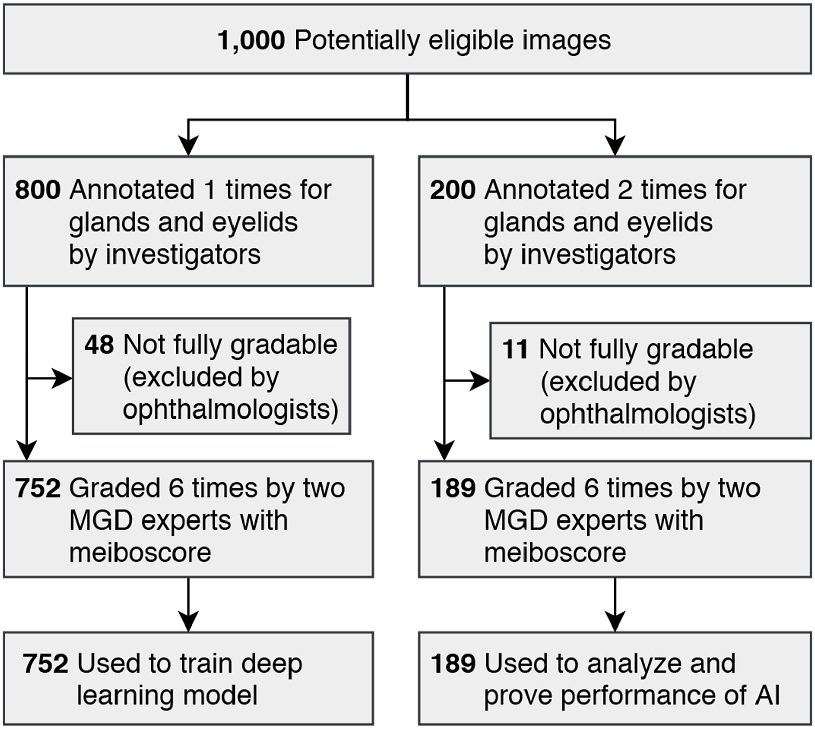
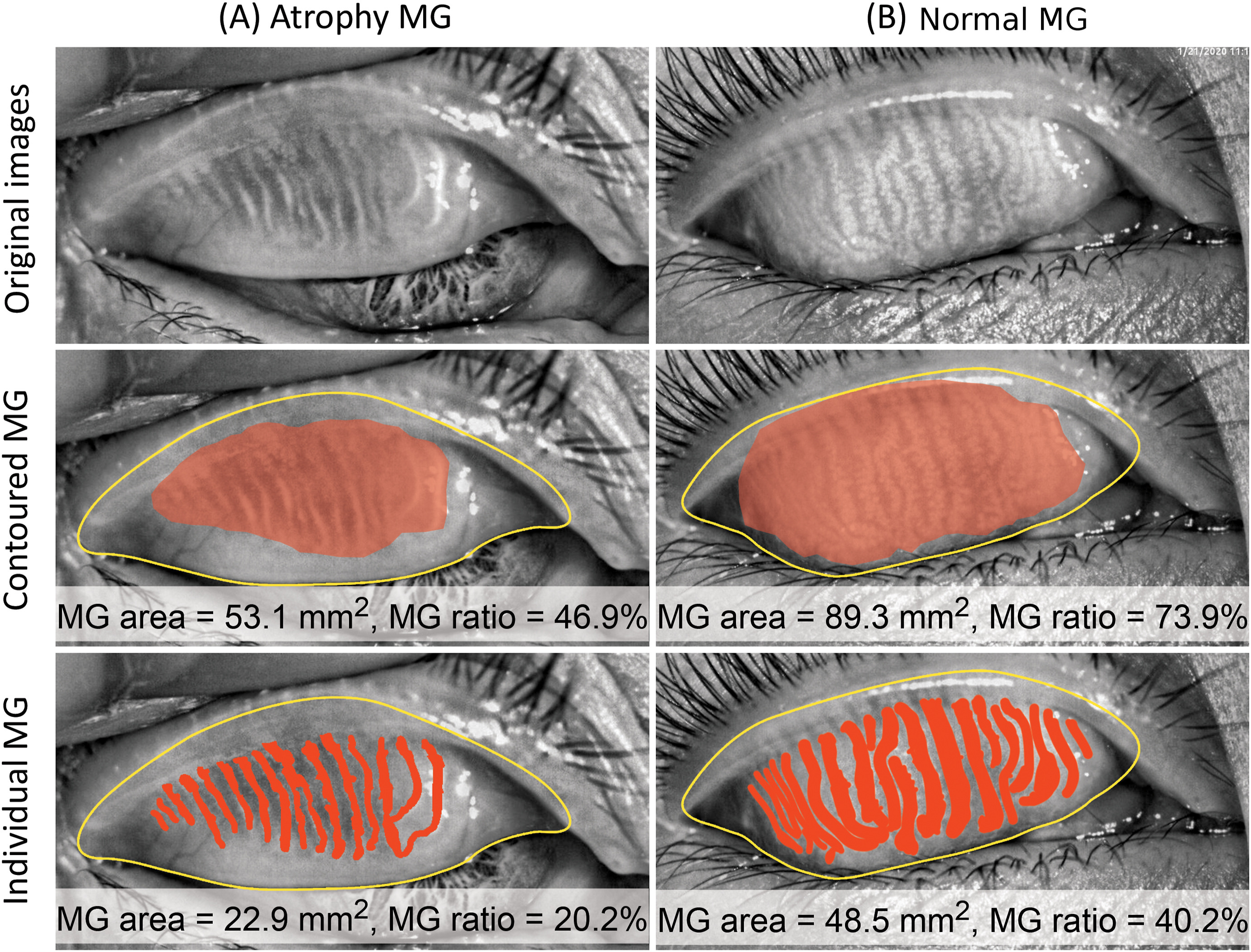
This MGD-1k dataset contains 1,000 infrared images of Meibomian Gland. All images are precisely annotated by investigators under direct supervision of MGD(meibomian gland dysfunction) experts and professional ophthalmologists.

This dataset is arranged into:-

  • 1000 images of Meibomian Gland
  • associated 1000 images of gland mask
  • associated 1000 images of eyelid mask
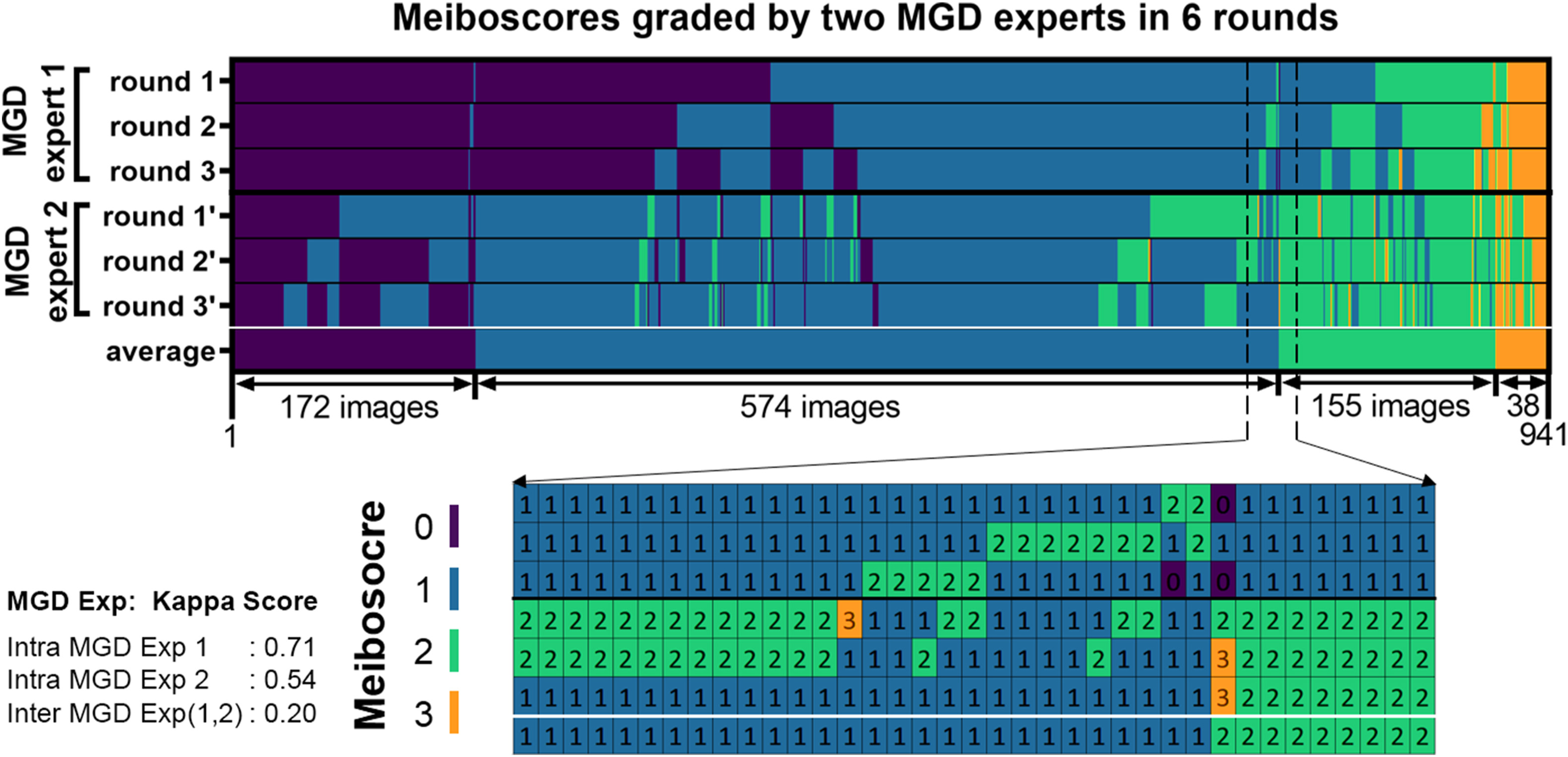
  • 6 rounds of meiboscore for each image

Dataset distribution

  • Number of Patients: 320
  • Age: Men (mean 51, std 19), Women (mean: 55 years,  std 19)
  • Men/Women ratio: 322(32.2%) vs 678 (67.8%)
  • Number of Meibomian Gland Images: 1000
  • Upper Eyelid images:  467
  • Lower Eyelid images:  533
  • Gradable Image* :  941 [94.1%]
  • Color Channel: Single/Grayscale  
  • Imaging Device: LipiView II Ocular Surface Interferometer
  • Duration of data collection: 2019 April to 2020 April (1 year)

* Within 1000 images, 59 images were marked as ungradable at least in one out of 6 rounds. All 941 images contain full gradings.

Demographic Distribution

Understanding MGD Across Age Groups

The MGD-1k dataset includes a diverse range of subjects, offering a substantial demographic representation for research into age-related patterns in Meibomian Gland Dysfunction. The age distribution of participants, as shown in the accompanying graph, provides critical insights into the prevalence and manifestation of MGD across different life stages.

Age Distribution of MG Patients
Dataset Curation

Annotation and Eligibility

The MGD-1k dataset was meticulously curated to ensure the highest quality. An extensive annotation process was followed by rigorous verification, which is critical for the dataset's utility in machine learning applications. This flowchart illustrates the detailed steps undertaken to prepare the dataset.

Age Distribution of MG Patients
Expert Validation

Consistency of Meiboscores

The high level of expert agreement on meiboscore grading showcased in this dataset underscores the precision and reliability of the annotations. The consistent grading by MGD experts across multiple rounds provides a strong foundation for AI-based diagnostic model development.

Meiboscores Validation
Morphological Analysis

Normal vs. Atrophic MG

The MGD-1k dataset provides invaluable morphological insights into Meibomian Gland Dysfunction by offering a detailed comparison between normal and atrophic gland conditions. These annotated images are crucial for advancing our understanding of MGD and refining diagnostic criteria.

Age Distribution of MG Patients

Download MGD-1k Dataset

Download 1000 Infrared Image  Download 1000 MG Mask  Download 1000 eyelid Mask  Download Graded Meiboscore Download Full dataset 

Project Members, Institution, and References

Author: The project was carried out by Ripon Kumar Saha, A. M. Mahmud Chowdhury, Kyung-Sun Na, Gyu Deok Hwang, Hae-Gon Jeon, Ho Sik Hwang, Euiheon Chung.

Institute: GIST | Gwangju Institute of Science and Technology, The Catholic University of Korea, Yeouido St. Mary's Hospital, College of Medicine.

References of Paper: Automated quantification of meibomian gland dropout in infrared meibography using deep learning, The Ocular Surface 2022, CiteScore: 14.1.

Contact: ripon.ece@gmail.com

Citation:
@article{saha2022automated,
          title={Automated quantification of meibomian gland dropout in infrared meibography using deep learning},
          author={Saha, Ripon Kumar and Chowdhury, AM Mahmud and Na, Kyung-Sun and Hwang, Gyu Deok and Eom, Youngsub and Kim, Jaeyoung and Jeon, Hae-Gon and Hwang, Ho Sik and Chung, Euiheon},
          journal={The Ocular Surface},
          volume={26},
          pages={283--294},
          year={2022},
          publisher={Elsevier}
        }